Scientists have captured the highest-resolution images ever made of DNA, revealing previously invisible twisting and twisting behavior.
Deoxyribonucleic acid, also known as DNA, can be surprisingly active when stuffed and twisted into a cell, according to new Research published in Nature Communications. These hidden movements were revealed by computer simulations fed with the highest resolution images ever taken of a single DNA molecule. The new study exposes previously unseen behavior in the self-replicating molecule, and this research could eventually lead to the development of powerful new genetic therapies.
“Seeing is believing, but with something as small as DNA, seeing the spiral structure of the entire DNA molecule was extremely challenging,” said Alice Pyne, the paper’s lead author and materials scientist at the University of Sheffield, in a statement from the University. “The videos we have developed allow us to observe DNA twisting with a level of detail never before seen.”

Scientists have earlier used microscopes to gaze at DNA and its twisted ladder-like configuration, but these were limited to static images of the molecule. What scientists have not been able to see is how intense DNA coiling affects the double helix structure. To accomplish this, Pyne and her colleagues combined high-resolution atomic force microscopy (AFM) with molecular dynamics computer simulations, which revealed the meandering.
G / O Media can receive a commission
Long, well-organized strands of DNA are densely packed in our cells. As the new study shows, this results in surprisingly dynamic physical behavior.

Agnes Noy, a lecturer at the University of York and a co-author of the study, said the microscopy images and computer simulations matched so well that they increased the resolution of their experiments, allowing the team to “ track how each atom of the double helix of DNA dances. “
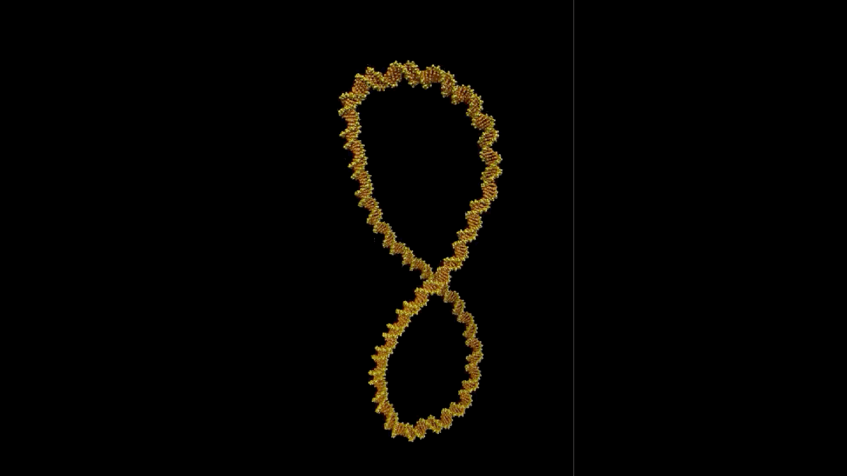
For the study, the researchers analyzed DNA mini-circles, in which a small strand is joined at both ends to form a loop structure. DNA mini-circles have been described before and are thought to be important health indicators.
Microscopic images of DNA mini-circles in their “relaxed” position (ie no twists) showed very little movement, but additional twists brought the loop to life, resulting in more powerful movements. These dynamic movements can serve an important purpose, helping the DNA find binding partners and facilitate growth.
The new atomic force microscopy shows, “with remarkable detail,” how “wrinkled, bubbly, kinked, denatured, and oddly shaped” the DNA mini-circles really are, “that we hope to one day be able to master,” Baylor College of Medicine biologist Lynn Zechiedrich, who provided the minicircles for the study, the University of Sheffield statement said.
Indeed, more insight into DNA and how it can become so compact could lead to the development of completely new medical interventions, including improved DNA-based diagnostics and therapeutics, according to the researchers.