
Credit: Cambridge University
Researchers have identified the driving force behind a cellular process related to neurodegenerative conditions such as Parkinson’s disease and motor neurons.
In a study published today in Scientific progress, Researchers at the University of Cambridge show that tiny components in the cell are the biological engines behind effective protein production.
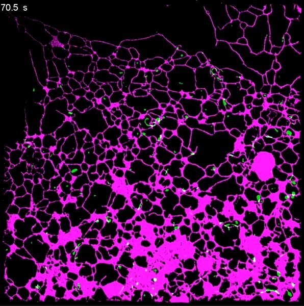
The endoplasmic reticulum (ER) is the protein factory of the cell and produces and modifies the proteins needed to ensure healthy cell function. It is the largest organelle in the cell and consists of a web-like structure of tubes and sheets. The ER moves quickly and changes shape constantly, extending across the cell to where it is needed at all times.
Using super-resolution microscopy techniques, researchers from Cambridge’s Department of Chemical Engineering and Biotechnology (CEB) have discovered the driving force behind these movements – a breakthrough that could have a significant impact on the study of neurodegenerative diseases.
“The endoplasmic reticulum is known to have a very dynamic structure – constantly stretching and expanding in the cell,” says Dr. Meng Lu, research associate at the Laser Analytics Group, led by Professor Clemens Kaminski.
“The ER must be able to reach all places efficiently and quickly to perform essential household tasks in the cell, when and where needed. Impairment of this ability has been linked to diseases such as Parkinson’s, Alzheimer’s, Huntington’s and ALS. has had a limited understanding of how the ER achieves these rapid and fascinating changes in shape and how it responds to cellular stimuli. “
Lu and colleagues found that another cell component holds the key: tiny structures that look like tiny droplets in membranes called lysosomes.
Lysosomes can be seen as the cell’s recycling centers: they trap damaged proteins and break them down to their original building blocks, so that they can be reused in the production of new proteins. Lysosomes also act as detection centers – they pick up environmental factors and communicate them with other parts of the cell, which adapt accordingly.
Up to 1000 lysosomes can fly through the cell at any one time, and with that, the ER appears to change its shape and location, in a seemingly orchestrated fashion.
What astonished the Cambridge scientists was their discovery of a causal relationship between the movement of the small lysosomes in the cell and the reforming process of the large ER network.
“We were able to show that it is the movement of the lysosomes themselves that forces the ER to reform in response to cellular stimuli,” said Lu. “When the cell feels that there is a need for lysosomes and ER to travel to the distal corners of the cell, the lysosomes pull the ER web with them, like little locomotives.”
From a biological point of view, this makes sense: the lysosomes act as a sensor in the cell and the ER as a response unit; Coordinating their synchronous function is critical to cellular health.
To discover this surprising bond between two very different organelles, Kaminski’s research team used new imaging technologies and machine learning algorithms, giving them unprecedented insight into the inner workings of the cell.
“It’s fascinating that we can now look inside living cells and see the tremendous speed and dynamics of the cellular machinery in such detail and in real time,” said Kaminski. “Just a few years ago it would have been unthinkable to see organelles going about their business in the cell.”
The researchers used lighting patterns projected onto living cells at high speed and advanced computer algorithms to recover information at a scale more than a hundred times the width of a human hair. It has only recently become possible to capture such information at video speeds.
The researchers also used machine learning algorithms to automatically extract the structure and movement of the ER networks and lysosomes from thousands of data sets.
The team expanded their research to look at neurons or nerve cells – specialized cells with long protrusions called axons, along which signals are sent. Axons are extremely thin tubular structures and it was not known how the motion of the very large ER network is orchestrated within these structures.
The study shows how lysosomes easily travel along the axons and drag the ER behind them. The researchers also show how disrupting this process is detrimental to the development of growing neurons.
Often the researchers saw events in which the lysosomes acted as repair engines for disconnected or broken pieces of ER structure, which they reassemble and fuse into an intact network. The work is therefore relevant to understanding nervous system disorders and their recovery.
The team also studied the biological significance of this coupled movement and provided an incentive – in this case, nutrients – for the lysosomes to perceive. The lysosomes appeared to move towards this signal, dragging the ER network after them so that the cell can elicit an appropriate response.
“Little was known so far about the regulation of ER structure in response to metabolic signals,” said Lu. “Our research links lysosomes as sensor units that actively direct the local ER response.”
The team hopes their insights will be invaluable to those studying the link between disease and cellular response, and their own next steps focus on studying ER function and dysfunction in diseases such as Parkinson’s and Alzheimer’s.
Neurodegenerative disorders have been associated with aggregation of damaged and misfolded proteins, so understanding the underlying mechanisms of ER function is critical for research on their treatment and prevention.
“The discoveries of the ER and lysosomes were awarded the Nobel Prize many years ago – they are essential organelles essential for healthy cellular function,” Kaminski said. “It is fascinating to think that there is still so much to learn about this system, which is incredibly important to the fundamental biomedical science that seeks the cause and cure for these devastating diseases.”
Lipids, Lysosomes, and Autophagy: The Keys to Preventing Kidney Damage
“The structure and worldwide distribution of the endoplasmic reticulum network is actively regulated by lysosomes” Science Advances (2020). advances.sciencemag.org/lookup… .1126 / sciadv.abc7209
Provided by University of Cambridge
Quote: Driving force behind cellular ‘protein factories’ identified (2020, December 16) Retrieved December 17, 2020 from https://phys.org/news/2020-12-cellular-protein-factories.html
This document is protected by copyright. Other than fair treatment for the purposes of private study or research, no part may be reproduced without written permission. The content is provided for informational purposes only.